Test Prep PCAT Pharmacy College Admission Test Online Training
Test Prep PCAT Online Training
The questions for PCAT were last updated at Nov 23,2024.
- Exam Code: PCAT
- Exam Name: Pharmacy College Admission Test
- Certification Provider: Test Prep
- Latest update: Nov 23,2024
A population of 1000 individuals has 110 births and 10 deaths in a year. Its growth rate (r) is equal to:
- A . 0.01 per year
- B . 0.11 per year
- C . 0.1 per year
- D . 0.09 per year
- E . 0.009 per year
C
Explanation:
The growth rate is equal to the difference between births and deaths divided by population size.
When a population reaches its carrying capacity?
- A . The population size begins to decrease.
- B . The population growth rate approaches zero.
- C . Other populations will be forced out of the habitat.
- D . Density-independent factors no longer play a role.
- E . Density-dependent factors no longer play a role.
B
Explanation:
Within a habitat, there is a maximum number of individuals that can continue to thrive, known as the habitat’s carrying capacity. When the population size approaches this number, population growth will stop.
Darwin’s idea that evolution occurs by the gradual accumulation of small changes can be summarized as:
- A . Convergent evolution
- B . Adaptive radiation
- C . Punctuated equilibrium
- D . Phyletic gradualism
- E . Sympatric speciation
D
Explanation:
Phyletic gradualism is the view that evolution occurs at a more or less constant rate. Contrary to this view, punctuated equilibrium holds that evolutionary history consists of long periods of stasis punctuated by geologically short periods of evolution. This theory predicts that there will be few fossils revealing intermediate stages of evolution, whereas phyletic gradualism views the lack of intermediate-stage fossils as a deficit in the fossil record that will resolve when enough specimens are collected.
At two independently assorting loci, a man has the following genotype: GgHH. He marries a woman with the genotype ggHh.
What is the probability that they will have a child who has the same genotype as the father?
- A . 0
- B . 1/2
- C . 1/4
- D . 1/8
C
Explanation:
This is a “probability” genetics question that can be answered by practical application of Mendel’s Laws. Mendel’s Law of Segregation states that alleles segregate during meiosis, resulting in gametes that carry only one allele for any given inherited trait (i.e., haploid gametes). Mendel’s Law of Independent Assortment states that unlinked genes assort independently during meiosis. By applying Mendel’s Laws, we can conclude that each parent in the problem can produce two possible gametes. The father can produce the gametes GH and gH, and the mother can produce the gametes gH and gh. The probability of the father’s genotype (GgHH) appearing in the progeny can be determined by calculating the number of different gamete combinations that will produce this genotype. Thus, a GgHH zygote can only be produced by the fusion of a GH gamete and a gH gamete. The probability that one parent will donate a particular gamete is independent of the probability that the other parent will donate a particular gamete. Thus, the probability of the father donating a GH gamete is 1/2, and the probability of the mother donating a gH gamete is 1/2. The probability of producing a genotype that requires the occurrence of both these independent events is equal to the product of the individual probabilities that these events will occur. Thus, 1/2 × 1/2 = 1/4, so the probability that this couple will have a child with the genotype GgHH is 1/4.
In a certain genetically stable population, the frequency of a recessive allele (for a trait with two alleles) is 0.6.
What is the frequency of individuals expressing the dominant trait?
- A . 0.64
- B . 0.36
- C . 0.24
- D . 0.16
A
Explanation:
The question stem asks you to determine the frequency of individuals expressing the dominant trait in a genetically stable population. However, before you do that, you need to determine the allelic frequencies in the population. This question involves a practical application of the Hardy-Weinberg equation. The Hardy-Weinberg equilibrium states that within a genetically stable population, the gene frequencies of dominant and recessive alleles will not change over time. Two mathematical expressions are associated with the Hardy-Weinberg equilibrium. The first relationship, p + q = 1, describes the relative allelic frequencies in a population. p is defined as the frequency of the dominant allele and q is defined as the frequency of the recessive allele, and the sum of both those frequencies adds up to 1, or 100%. The second relationship, p2 + 2pq+ q2 = 1, describes the relative genotypic frequencies in the population. p2 represents homozygous, or dominant pp genotypes; q2 represents homozygous, or frequency of the dominant allele, p, by the mathematical relationship p + q = 1. Therefore, the frequency of p is 0.4 because 0.6 + 0.4 = 1. Next, you need to determine the frequency of individuals expressing the dominant trait by recessive qq genotypes; and 2pq represents the frequency of heterozygotes, or hybrids.applying the second relationship, p2 + 2pq+ q2 = 1. The individuals expressing the dominant trait are those that have the pp and pq genotypes, so to find the total frequency of individuals expressing the dominant trait, you add p2 and 2pq. Thus, p2 = 0.4 × 0.4, or 0.16 and 2pq = 2 × 0.6 × 0.4, or 0.48. If you add the two together, you get 0.16 + 0.48, or 0.64. Thus, 0.64 is the correct frequency of individuals expressing the dominant trait.
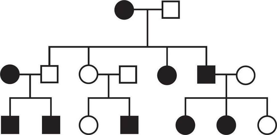
What is the inheritance pattern of the observed trait indicated by the pedigree below?

- A . Autosomal recessive
- B . Autosomal dominant
- C . X-linked recessive
- D . X-linked
- E . Cannot be determined
A
Explanation:
Pedigrees show the distribution of a single observable trait, or phenotype, across a family tree. In classical genetics, each phenotype is determined by a combination of two alleles contributed by two copies of the same (but not necessarily identical) chromosome. One allele is generally dominant, meaning it is expressed if it is present at all. In contrast, the other allele is recessive, meaning it is only expressed in the absence of a dominant allele, which generally means two copies need to be inherited to display the recessive phenotype. The exceptions are those alleles found on the X chromosome in males; males’ sex chromosomes include only one X (and one Y), so each trait coded for on the X chromosome is determined by only one allele instead of a combination of two alleles. This means it’s statistically more likely for males to inherit recessive X-linked traits since only one copy of the recessive alleles needs to be inherited to display the recessive phenotypes, as opposed to the usual two.
The fastest way to determine which inheritance pattern is shown by a pedigree, then, is to use the Kaplan shortcut: Identify whether two matching parents have an opposite offspring. If two affected parents have an unaffected offspring, both parents must have been heterozygous (having one of each allele), and the trait must be dominant: Rr × Rr C> rr. If two unaffected parents have an affected offspring, both parents must have one again been heterozygous, but in that situation, the trait being tracked must have been the recessive one: Rr × Rr C> rr. In the pedigree provided in this question, generational skipping occurs in the middle portion: Generation 2 has two unaffected parents, but generation 3 has an affected offspring. This indicates a recessive trait. Since a roughly equal number of males and females are affected (5 : 4 ratio), this is an autosomal trait.
When blood flow to human tissue is interrupted, the lack of sufficient blood supply is called ischemia. If ischemia is not restored quickly, the affected tissue may undergo a process called infarction, which involves a series of chemical changes that damage the tissue. The lack of blood supply results in lack of oxygen, and thus lactic acidosis. Mitochondrial dysfunction results. Microscopic examination and chemical analysis of ischemic cells reveal membrane degeneration, excessive calcium (Ca+) inside the cell, and free radical formation, accompanied by a reactive inflammation and free fatty acid formation. A research experiment is designed to evaluate the response of infarcted tissue to intra-arterial administration of an antioxidant. Preliminary results demonstrate that follow-up evaluation of tissue exposed to intra-arterial antioxidant injection resulted, on average, in a smaller area of infarcted tissue after seven days when compared to controls without exposure to the antioxidant. It was noted that 70% of the patients who demonstrated smaller areas of infarction also had a notable decease in edema of the ischemic tissue lasting about 6 to 10 hours after injection.
What is a possible explanation for the relationship among antioxidant injection, edema, and tissue damage?
- A . Antioxidants produce anti-infarction biochemical reactions that decrease the size of the infarct.
- B . Antioxidants decrease tissue damage by decreasing edema.
- C . The prevention of tissue damage may be produced by a combination of the effect of decreased edema and the injection of antioxidants.
- D . Increased blood flow causes paradoxical tissue damage due to ischemia.
C
Explanation:
The experimental results do not demonstrate or prove that the antioxidant is responsible for the decrease in edema or that edema is the cause of tissue damage. However, because patients exposed to the antioxidant had a smaller area of infarcted tissue, it appears that the antioxidant has a beneficial effect. Most, but not all, of the patients with smaller areas of infarct also had decreased edema, suggesting that edema may also play a role. This suggests that some type of combination of the presence of edema and antioxidants was at play when decreased tissue damage was observed. There was no measured relationship to blood flow. It is unclear exactly why the antioxidant injected samples showed deceased damage, and it is a leap to suggest that the antioxidants themselves produce chemicals or biochemical reactions that decreased the size of the infarct or the edema.
When blood flow to human tissue is interrupted, the lack of sufficient blood supply is called ischemia. If ischemia is not restored quickly, the affected tissue may undergo a process called infarction, which involves a series of chemical changes that damage the tissue. The lack of blood supply results in lack of oxygen, and thus lactic acidosis. Mitochondrial dysfunction results. Microscopic examination and chemical analysis of ischemic cells reveal membrane degeneration, excessive calcium (Ca+) inside the cell, and free radical formation, accompanied by a reactive inflammation and free fatty acid formation. A research experiment is designed to evaluate the response of infarcted tissue to intra-arterial administration of an antioxidant. Preliminary results demonstrate that follow-up evaluation of tissue exposed to intra-arterial antioxidant injection resulted, on average, in a smaller area of infarcted tissue after seven days when compared to controls without exposure to the antioxidant. It was noted that 70% of the patients who demonstrated smaller areas of infarction also had a notable decease in edema of the ischemic tissue lasting about 6 to 10 hours after injection.
How could lactic acid production and free fatty acid formation contribute to organelle dysfunction?
- A . The acidity of these molecular products, when uncorrected, alters the cell’s pH beyond that which the cell can compensate for. Organelles, containing proteins, denature as a result.
- B . Lactic acid production and free fatty acid formation function like free radicals, altering the structure of the molecular components of the organelles.
- C . Lactic acids and free fatty acids crowd the organelles within the cells, preventing them from communicating with each other in the cytoplasm.
- D . Lactic acids and free fatty acids are hydrophobic and thus can enter the membranes of the organelles, disrupting their function.
A
Explanation:
In small quantities, lactic acids and free fatty acids are tolerable due to the cell’s ability to buffer mild pH changes. However, in an ischemic setting, the cell cannot correct the pH changes, and thus the proteins that form the structural and functional components of the organelles begin to denature. Lactic acids and free fatty acids are acidic, meaning that they contribute hydrogen atoms to the environment, whereas free radicals are deficient in electrons. Although the volume of acidic molecules within the cell is not beneficial for the organelles, their pH is their most harmful characteristic and thus the most immediately damaging consequence. Free fatty acids are hydrophobic and thus may be able to pass through organelle membranes, but they cause organelle dysfunction from outside the organelle in the cytoplasm as well.
When blood flow to human tissue is interrupted, the lack of sufficient blood supply is called ischemia. If ischemia is not restored quickly, the affected tissue may undergo a process called infarction, which involves a series of chemical changes that damage the tissue. The lack of blood supply results in lack of oxygen, and thus lactic acidosis. Mitochondrial dysfunction results. Microscopic examination and chemical analysis of ischemic cells reveal membrane degeneration, excessive calcium (Ca+) inside the cell, and free radical formation, accompanied by a reactive inflammation and free fatty acid formation. A research experiment is designed to evaluate the response of infarcted tissue to intra-arterial administration of an antioxidant. Preliminary results demonstrate that follow-up evaluation of tissue exposed to intra-arterial antioxidant injection resulted, on average, in a smaller area of infarcted tissue after seven days when compared to controls without exposure to the antioxidant. It was noted that 70% of the patients who demonstrated smaller areas of infarction also had a notable decease in edema of the ischemic tissue lasting about 6 to 10 hours after injection.
Which of the following chemical moieties forms the backbone of DNA?
- A . Nitrogenous bases
- B . Glycerol
- C . Amino groups
- D . Pentose and phosphate
D
Explanation:
DNA is composed of nucleotides joined together in long chains. Nucleotides are composed of a pentose sugar, a phosphate group, and a nitrogenous base. The bases form the “rungs” of the ladder at the core of the DNA helix and the pentose-phosphates are on its outside, or backbone.
Which of the following organelles helps green plants synthesize organic compounds like starch in the presence of sunlight?
- A . Mitochondria
- B . Chloroplast
- C . Ribosomes
- D . Golgi body
B
Explanation:
Green plants, with the help of sunlight and in the presence of enzymes, synthesize organic compounds like starch from inorganic compounds like CO 2 and H 2 O. This is known as photosynthesis. Chloroplast is the organelle to perform photosynthesis. Plants that are devoid of chloroplast cannot synthesize starch.
Latest PCAT Dumps Valid Version with 282 Q&As
Latest And Valid Q&A | Instant Download | Once Fail, Full Refund

